
Lumbar facet syndrome is a clinical diagnosis referring to low back pain caused by injury or deterioration of facet joints in the lumbar spine. We discuss
WHAT IS LUMBAR FACET SYNDROME ?
Facet syndrome is a clinical diagnosis referring to low back pain or neck pain caused by injury or deterioration of facet joint in the lumbar or cervical spine. Before we explore causes and treatment options for this problem, let me briefly review what facet joint is, and what is its function.
FUNCTIONAL SPINAL UNIT
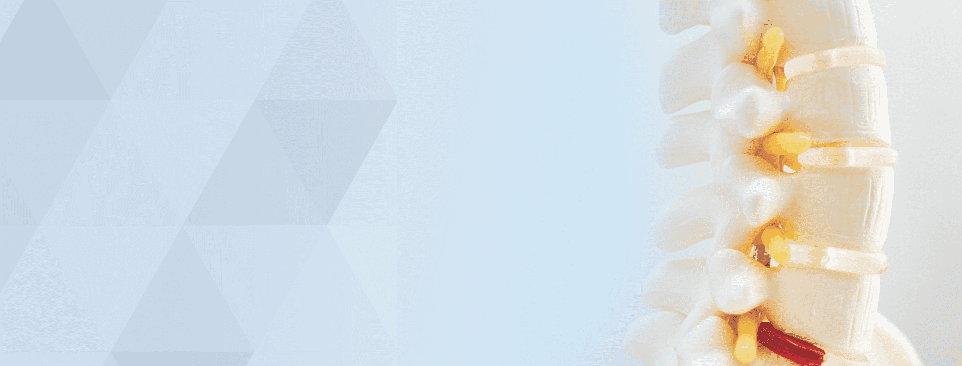
The human spine consists of several vertebrae that are connected to each other via 3 joints. In the front portion of the human spine, there is intervertebral disc, which provides cushioning of compression forces and transfer the energy along the spine. On the back side of the spine, there is a pair of smaller facet joints that contribute to the spine motion and govern torsional movements at given level (Fig.1) The intervertebral disc and 2 facet joints are responsible for transfer energy from spine to legs and allow humans to move in upright fashion. The 2 adjacent vertebrae separated by the intervertebral disc and 2 facet joints represent one functional spinal unit.

FACET JOINT ANATOMY
The facet joint consists of two opposing bony processes from the adjacent vertebrae. Each process has small contact area that is covered by the cartilage. The both processes are wrapped in joint capsule. The capsule provides joint stability and holds the synovial fluid that lubricate joint surfaces. The cartilage in healthy facet joint is smooth like billiard ball allowing friction-less motion. Figure 2 depicts healthy facet joint. As we age or in a case of facet injury, the cartilage wears off and the surface become rough. The capsule will also get loose or damaged and irritated. We refer to these changes as arthritic remodeling. Figure 2 also depicts damaged facet joint. The cartilage is worn down. There are several bony osteophytes (bumps) around the periphery of the joint surface limiting facet joint motion. The joint is often irritated and inflamed as demonstrated by redness.

FACET JOINT INNERVATION
Facet joint represents very important structure allowing human locomotion. The capsule contains several sensory (pain sensing) nerve fibers that joins bigger spinal nerve roots and carry sensory and pain signals to patient’s brain. Figure 3 depicts model of facet joint with red capsule. There are several yellow fibers that represent sensory nerves. We refer to them as median branches. The Figure 3 also shows how these nerves run towards bigger spinal nerve roots.

This is very important anatomic relationship as this helps physician decipher patient’s symptoms and understand source of the pain. Here is what is likely going on in physician’s brain as he/she listens to the patient complaints. If a facet joint has been injured, subsequent inflammation irritates nerve fibers in the capsule. Logically the patient feels pain in affected area. In addition, CNS will try to protect affected spine segment by signaling muscles in affected area to limit the motion of damaged segment. Again, the patient will perceive this as muscle spasms or stiffness in lumbar or cervical area. The typical compliant we hear from the patients suffering from facet syndrome, is the progressively worsening pain with prolong standing. This is again very logical as upright posture increases pressure on facet joints causing the aggravation of the pain. Furthermore, the patient often describe radiation of pain to buttocks or thighs. This is again very logical as sensory fibers run to bigger spinal nerve roots. These bigger nerves may also get irritated. The end result is that the patient feels pain spreading (radiating) to buttocks or upper thighs.
HOW IS FACET SYNDROME DIAGNOSED?
For experienced physicians these symptoms are very logical and clear. This may seem as just another exercise in functional anatomy. However, experienced physician seldom stop there. S/he needs to prove his/her suspicion before suggesting a specific treatment plan. If the facet is prime suspect, the physician may ask a patient to bend backwards and to the side. This maneuver will increase pressure in the affected facet joint and the patient will likely report aggravation of the pain. If this is the case, we can proceed to last step in diagnostic process.

local anesthetic to proximity of the median branch nerve to diagnose source of the pain.
No reasonable physician would intentionally aggravate patient’s symptoms and then leave him/her suffering. Therefore, it is prudent to finish our diagnostic process with performing a nerve block. This is a relatively simple (not easy) procedure which goal is to inject a small amount of local anesthetic agent close to suspected facet joint. Similarly to dentist who may knock on patient’s teeth to find aching tooth and subsequently numb it, we also will place needle close to suspected facet join and inject numbing medicine. Figure 4 shows a needle placed at outer side of left L4 facet joint and injection of local anesthetic ( magenta color ) to numb median nerve branches.
In some cases, physician will inject mixture of local anesthetic with steroid that may suppress inflammation and provide longer lasting pain relief. Unfortunately, the pain tends to return after some time and nobody really can predict how long the pain relief will last. The nerve block is a diagnostic procedure and should not be used repeatedly as a treatment.
WHAT ARE TREATMENT OPTIONS?
If the block provided significant pain relief (more than 75% reduction of the pain), but the effect did not last long enough (only few days), we can permanently desensitize the facet joint by destroying sensory nerve branches using either radiofrequency or endoscopic laser procedure. These procedures are very similar to root canal treatment in dentistry during which a dentist removes sensory nerves and seal the aching tooth. Radiofrequency ablation and endoscopic laser procedure are not the same. They have significant differences that are beyond scope of this article. We will explain these in separate article as well as address other relevant treatment options.
